Periodontitis lizards Dental
![]()
![]()
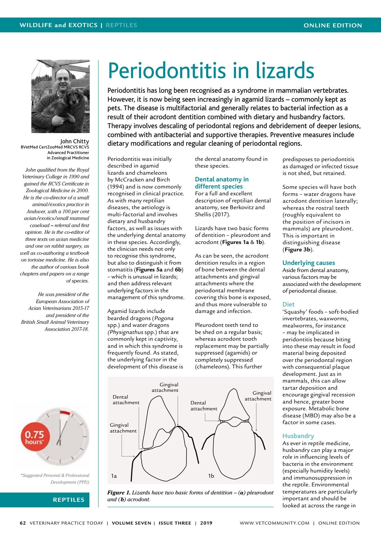
ONLINE EDITIONWILDLIFE and EXOTICS | REPTILES VETERINARY PRACTICE TODAY | VOLUME SEVEN | ISSUE THREE | 2019 62 REPTILES Suggested Personal & Professional Development (PPD) John Chitty BVetMed CertZooMed MRCVS RCVS Advanced Practitioner in Zoological Medicine John qualified from the Royal Veterinary College in 1990 and gained the RCVS Certificate in Zoological Medicine in 2000. He is the co-director of a small animal/exotics practice in Andover, with a 100 per cent avian/exotics/small mammal caseload referral and first opinion. He is the co-editor of three texts on avian medicine and one on rabbit surgery, as well as co-authoring a textbook on tortoise medicine. He is also the author of various book chapters and papers on a range of species. He was president of the European Association of Avian Veterinarians 2015-17 and president of the British Small Animal Veterinary Association 2017-18. Periodontitis in lizards Periodontitis has long been recognised as a syndrome in mammalian vertebrates. However, it is now being seen increasingly in agamid lizards commonly kept as pets. The disease is multifactorial and generally relates to bacterial infection as a result of their acrodont dentition combined with dietary and husbandry factors. Therapy involves descaling of periodontal regions and debridement of deeper lesions, combined with antibacterial and supportive therapies. Preventive measures include dietary modifications and regular cleaning of periodontal regions. Periodontitis was initially described in agamid lizards and chameleons by McCracken and Birch (1994) and is now commonly recognised in clinical practice. As with many reptilian diseases, the aetiology is multi-factorial and involves dietary and husbandry factors, as well as issues with the underlying dental anatomy in these species. Accordingly, the clinician needs not only to recognise this syndrome, but also to distinguish it from stomatitis ( Figures 5a and 6b ) which is unusual in lizards; and then address relevant underlying factors in the management of this syndrome. Agamid lizards include bearded dragons (Pogona spp.) and water dragons (Physignathus spp.) that are commonly kept in captivity, and in which this syndrome is frequently found. As stated, the underlying factor in the development of this disease is the dental anatomy found in these species. Dental anatomy in different species For a full and excellent description of reptilian dental anatomy, see Berkovitz and Shellis (2017). Lizards have two basic forms of dentition pleurodont and acrodont ( Figures 1a & 1b ). As can be seen, the acrodont dentition results in a region of bone between the dental attachments and gingival attachments where the periodontal membrane covering this bone is exposed, and thus more vulnerable to damage and infection. Pleurodont teeth tend to be shed on a regular basis; whereas acrodont tooth replacement may be partially suppressed (agamids) or completely suppressed (chameleons). This further predisposes to periodontitis as damaged or infected tissue is not shed, but retained. Some species will have both forms water dragons have acrodont dentition laterally; whereas the rostral teeth (roughly equivalent to the position of incisors in mammals) are pleurodont. This is important in distinguishing disease ( Figure 3b ). Underlying causes Aside from dental anatomy, various factors may be associated with the development of periodontal disease. Diet Squashy foods soft-bodied invertebrates, waxworms, mealworms, for instance may be implicated in peridontitis because biting into these may result in food material being deposited over the periodontal region with consequential plaque development. Just as in mammals, this can allow tartar deposition and encourage gingival recession and hence, greater bone exposure. Metabolic bone disease (MBD) may also be a factor in some cases. Husbandry As ever in reptile medicine, husbandry can play a major role in influencing levels of bacteria in the environment (especially humidity levels) and immunosuppression in the reptile. Environmental temperatures are particularly important and should be looked at across the range in Figure 1. Lizards have two basic forms of dentition ( a ) pleurodont and ( b ) acrodont. Dental attachment Gingival attachment Gingival attachment Dental attachment Gingival attachment 1a 1b WWW.VETCOMMUNIT Y.COM | ONLINE EDITION