Treatment ONLINE EDITION
![]()
![]()
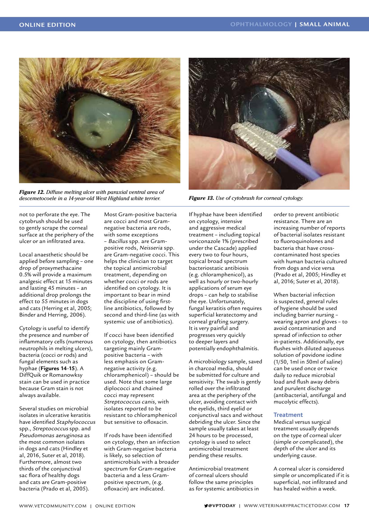
ONLINE EDITION OPHTHALMOLOGY | SMALL ANIMAL VPTODAY | WWW.VETERINARYPRACTICETODAY.COM 17 not to perforate the eye. The cytobrush should be used to gently scrape the corneal surface at the periphery of the ulcer or an infiltrated area. Local anaesthetic should be applied before sampling one drop of proxymethacaine 0.5% will provide a maximum analgesic effect at 15 minutes and lasting 45 minutes an additional drop prolongs the effect to 55 minutes in dogs and cats (Herring et al, 2005; Binder and Herring, 2006). Cytology is useful to identify the presence and number of inflammatory cells (numerous neutrophils in melting ulcers), bacteria (cocci or rods) and fungal elements such as hyphae ( Figures 14 - 15 ). A DiffQuik or Romanowksy stain can be used in practice because Gram stain is not always available. Several studies on microbial isolates in ulcerative keratitis have identified Staphylococcus spp., Streptococcus spp. and Pseudomonas aeruginosa as the most common isolates in dogs and cats (Hindley et al, 2016, Suter et al, 2018). Furthermore, almost two thirds of the conjunctival sac flora of healthy dogs and cats are Gram-positive bacteria (Prado et al, 2005). Most Gram-positive bacteria are cocci and most Gram- negative bacteria are rods, with some exceptions Bacillus spp. are Gram- positive rods, Neisseria spp. are Gram-negative cocci. This helps the clinician to target the topical antimicrobial treatment, depending on whether cocci or rods are identified on cytology. It is important to bear in mind the discipline of using first- line antibiotics, followed by second and third-line (as with systemic use of antibiotics). If cocci have been identified on cytology, then antibiotics targeting mainly Gram- positive bacteria with less emphasis on Gram- negative activity (e.g. chloramphenicol) should be used. Note that some large diplococci and chained cocci may represent Streptococcus canis, with isolates reported to be resistant to chloramphenicol but sensitive to ofloxacin. If rods have been identified on cytology, then an infection with Gram-negative bacteria is likely, so selection of antimicrobials with a broader spectrum for Gram-negative bacteria and a less Gram- positive spectrum, (e.g. ofloxacin) are indicated. If hyphae have been identified on cytology, intensive and aggressive medical treatment including topical voriconazole 1% (prescribed under the Cascade) applied every two to four hours, topical broad spectrum bacteriostatic antibiosis (e.g. chloramphenicol), as well as hourly or two-hourly applications of serum eye drops can help to stabilise the eye. Unfortunately, fungal keratitis often requires superficial keratectomy and corneal grafting surgery. It is very painful and progresses very quickly to deeper layers and potentially endophthalmitis. A microbiology sample, saved in charcoal media, should be submitted for culture and sensitivity. The swab is gently rolled over the infiltrated area at the periphery of the ulcer, avoiding contact with the eyelids, third eyelid or conjunctival sacs and without debriding the ulcer. Since the sample usually takes at least 24 hours to be processed, cytology is used to select antimicrobial treatment pending these results. Antimicrobial treatment of corneal ulcers should follow the same principles as for systemic antibiotics in order to prevent antibiotic resistance. There are an increasing number of reports of bacterial isolates resistant to fluoroquinolones and bacteria that have cross- contaminated host species with human bacteria cultured from dogs and vice versa (Prado et al, 2005; Hindley et al, 2016; Suter et al, 2018). When bacterial infection is suspected, general rules of hygiene should be used including barrier nursing wearing apron and gloves to avoid contamination and spread of infection to other in-patients. Additionally, eye flushes with diluted aqueous solution of povidone iodine (1/50, 1ml in 50ml of saline) can be used once or twice daily to reduce microbial load and flush away debris and purulent discharge (antibacterial, antifungal and mucolytic effects). Treatment Medical versus surgical treatment usually depends on the type of corneal ulcer (simple or complicated), the depth of the ulcer and its underlying cause. A corneal ulcer is considered simple or uncomplicated if it is superficial, not infiltrated and has healed within a week. Figure 12. Diffuse melting ulcer with paraxial ventral area of descemetocoele in a 14-year-old West Highland white terrier. Figure 13. Use of cytobrush for corneal cytology. WWW.VETCOMMUNIT Y.COM | ONLINE EDITION