Treatment simple superficial
![]()
![]()
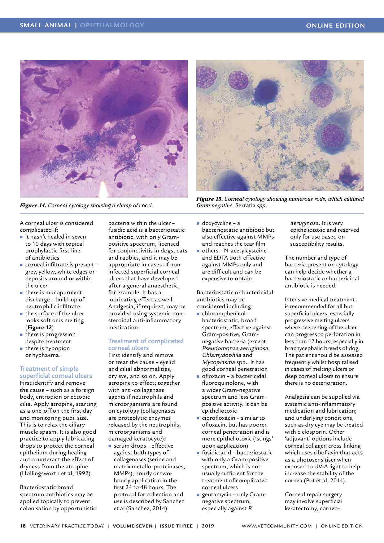
ONLINE EDITIONSMALL ANIMAL | OPHTHALMOLOGY VETERINARY PRACTICE TODAY | VOLUME SEVEN | ISSUE THREE | 2019 18 A corneal ulcer is considered complicated if: it hasnt healed in seven to 10 days with topical prophylactic first-line of antibiotics corneal infiltrate is present grey, yellow, white edges or deposits around or within the ulcer there is mucopurulent discharge build-up of neutrophilic infiltrate the surface of the ulcer looks soft or is melting ( Figure 12 ) there is progression despite treatment there is hypopion or hyphaema. Treatment of simple superficial corneal ulcers First identify and remove the cause such as a foreign body, entropion or ectopic cilia. Apply atropine, starting as a one-off on the first day and monitoring pupil size. This is to relax the ciliary muscle spasm. It is also good practice to apply lubricating drops to protect the corneal epithelium during healing and counteract the effect of dryness from the atropine (Hollingsworth et al, 1992). Bacteriostatic broad spectrum antibiotics may be applied topically to prevent colonisation by opportunistic bacteria within the ulcer fusidic acid is a bacteriostatic antibiotic, with only Gram- positive spectrum, licensed for conjunctivitis in dogs, cats and rabbits, and it may be appropriate in cases of non- infected superficial corneal ulcers that have developed after a general anaesthetic, for example. It has a lubricating effect as well. Analgesia, if required, may be provided using systemic non- steroidal anti-inflammatory medication. Treatment of complicated corneal ulcers First identify and remove or treat the cause eyelid and cilial abnormalities, dry eye, and so on. Apply atropine to effect; together with anti-collagenase agents if neutrophils and microorganisms are found on cytology (collagenases are proteolytic enzymes released by the neutrophils, microorganisms and damaged keratocyte): serum drops effective against both types of collagenases (serine and matrix metallo-proteinases, MMPs), hourly or two- hourly application in the first 24 to 48 hours. The protocol for collection and use is described by Sanchez et al (Sanchez, 2014). doxycycline a bacteriostatic antibiotic but also effective against MMPs and reaches the tear film others N-acetylcysteine and EDTA both effective against MMPs only and are difficult and can be expensive to obtain. Bacteriostatic or bactericidal antibiotics may be considered including: chloramphenicol bacteriostatic, broad spectrum, effective against Gram-positive, Gram- negative bacteria (except Pseudomonas aeruginosa, Chlamydophila and Mycoplasma spp.. It has good corneal penetration ofloxacin a bactericidal fluoroquinolone, with a wider Gram-negative spectrum and less Gram- positive activity. It can be epitheliotoxic ciprofloxacin similar to ofloxacin, but has poorer corneal penetration and is more epitheliotoxic (stings upon application) fusidic acid bacteriostatic with only a Gram-positive spectrum, which is not usually sufficient for the treatment of complicated corneal ulcers gentamycin only Gram- negative spectrum, especially against P. aeruginosa. It is very epitheliotoxic and reserved only for use based on susceptibility results. The number and type of bacteria present on cytology can help decide whether a bacteriostatic or bactericidal antibiotic is needed. Intensive medical treatment is recommended for all but superficial ulcers, especially progressive melting ulcers where deepening of the ulcer can progress to perforation in less than 12 hours, especially in brachycephalic breeds of dog. The patient should be assessed frequently whilst hospitalised in cases of melting ulcers or deep corneal ulcers to ensure there is no deterioration. Analgesia can be supplied via systemic anti-inflammatory medication and lubrication; and underlying conditions, such as dry eye may be treated with ciclosporin. Other adjuvant options include corneal collagen cross-linking which uses riboflavin that acts as a photosensitiser when exposed to UV-A light to help increase the stability of the cornea (Pot et al, 2014). Corneal repair surgery may involve superficial keratectomy, corneo- Figure 14. Corneal cytology showing a clump of cocci. Figure 15. Corneal cytology showing numerous rods, which cultured Gram-negative, Serratia spp.. WWW.VETCOMMUNIT Y.COM | ONLINE EDITION